VITREOMACULAR TRACTION

How Does Vitreomacular Traction Occur?
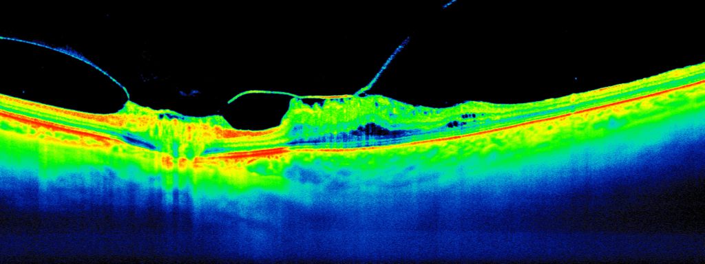
Vitreomacular traction occurs when the vitreous jelly inside the eye pulls or exerts a force on the macula. This tractional force, generated by the adhesion of the vitreous to the macula, distorts the normal contour of the retina. It results in blurred and/or distorted vision. Similar symptoms are also experienced in other conditions affecting the macula, such as age-related macular degeneration and epiretinal membrane.
Why Does Traction On the Macula Occur?
The vitreous jelly is normally firmly attached to the retina but it becomes more watery with age. The vitreous may eventually detach from the retina in a process known as posterior vitreous detachment. However, if the vitreous jelly does not fully separate from the retina, leaving points of contact or adhesions, the remaining partially attached vitreous can pull and distort the macula. Consequently, this results in vitreomacular traction. The condition is best diagnosed using high-resolution optical coherence tomography.
What Are the Treatment Options?
If you do not have any symptoms in your vision from the vitreomacular traction, we recommend observation and regular monitoring. Additionally, we advise that you check your vision at home regularly using an Amsler grid and look out for any changes in vision or new symptoms. Some cases of vitreomacular traction syndrome resolve spontaneously without treatment.
Vitrectomy
However, if you experience significant blurred or distorted vision, Dr Lee may recommend vitrectomy surgery help relieve the pulling or traction on the macula and restore your vision.
The success rate of vitrectomy is high. Patients experience significant improvement in vision. Dr Lee will discuss the benefits and risks of surgery and provide instructions regarding post-operative recovery.
