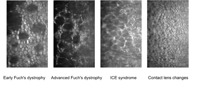
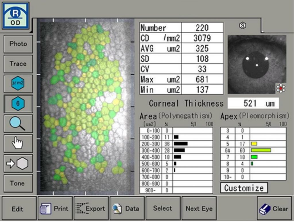
The endothelium is a single layer of cells on the inner surface of the cornea. It specifically functions to maintain the clarity of the cornea. Diseases of the endothelium cause swelling of the cornea resulting in opacity, loss of vision and breakdown of the eye surface resulting in ulceration and scarring.
At City Eye Centre, we are able to assess and monitor the structure of the endothelium using specular microscopy. This specialised equipment is able to assess the size, shape and density of the cells as well as identifying any abnormal structures. Important diseases of the endothelium include Fuch’s endothelial dystrophy, iridocorneal endothelial syndrome (ICE), contact lenses induced changes, posterior polymorphous dystrophy and endothelial cell loss due to previous surgery or trauma. Specular microscopy is also an important monitoring tool post corneal transplantation.


